
Book a Consultation
Thank you!
Your form has been sent successfully.


January 13, 2024
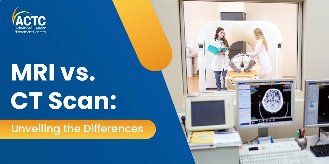
Magnetic Resonance Imaging (MRI) and Computed Tomography (CT) scans are two imaging modalities that stand out as essential components of contemporary healthcare.
This technology has completely transformed healthcare practitioners' understanding and visualization of the human body. Even though they are both formidable devices, they have distinct uses and qualities that make them appropriate in particular circumstances.
MRI stands for magnetic resonance imaging, a non-invasive imaging method that produces finely detailed images of the body's internal components using strong magnets and radio waves.
Since MRIs don't employ ionizing radiation like CT scans or X-rays do, they are a safer option for certain patient populations, such as younger patients and pregnant women.
The behavior of hydrogen atoms in the body in the presence of a high magnetic field is the basic idea underlying magnetic resonance imaging (MRI). These atoms are aligned by the magnetic field, and this alignment is then momentarily disturbed by radiofrequency pulses.
The MRI equipment picks up the signals that the atoms release as they realign, producing intricate cross-sectional pictures of the region being studied.
In contrast, X-rays are used in Computed Tomography or CT scan, an imaging method that creates finely detailed, cross-sectional images of the body. For concurrent examination of soft tissues, organs, and bones, CT scans are especially helpful.
The technique of fusing several X-ray pictures to get a comprehensive, three-dimensional picture of the inside structures is known as "computed tomography".
The patient lies on a table that passes through the CT machine's circular aperture to undergo a CT scan. Various angles are used to send X-ray beams through the body; once they pass through, the X-rays are detected by detectors on the other side of the apparatus.
After processing this data, a computer creates intricate cross-sectional pictures, or slices, of the body.
Nuclear magnetic resonance serves as the foundation for MRI technology. It depends on how radiofrequency pulses interact with the body's hydrogen nuclei's magnetic characteristics. The quality and resolution of the photographs are significantly influenced by the magnetic field's strength, which is expressed in Tesla (T). In general, photographs with stronger magnetic fields are sharper and more detailed.
MRI equipment comes in two varieties: open MRIs and closed, or classic, MRIs. Open MRI scanners offer a more open design, making them more suitable for people who might have claustrophobia. On the other hand, closed MRI machines—which resemble tunnels—are frequently more potent and yield images with greater resolution.
The core of CT scan technology is X-ray imaging. Within the CT machine, a rotating gantry holds the detectors and X-ray tube. The X-ray beam spins around the patient's body as they pass through the circular hole, taking pictures from various perspectives. These photos are processed by the computer to produce intricate cross-sectional views.
Differentiating between distinct body structures is based on the amount of X-rays absorbed, which is influenced by the density of the tissues. Modern CT scanners are useful in emergency situations where a prompt and precise diagnosis is essential since they can create images with incredibly fine detail and offer short scanning times.
MRI is renowned for its ability to provide detailed images of soft tissues and is often the preferred imaging modality for the brain, spinal cord, joints, and organs such as the heart and liver.
Its sensitivity to changes in tissue composition makes it a valuable tool for detecting abnormalities, such as tumors, inflammation, and structural abnormalities.
Neuroimaging: MRI is the primary tool for imaging the brain and spinal cord. It is highly effective in detecting abnormalities such as brain tumors, vascular malformations, and degenerative diseases.
Musculoskeletal Imaging: MRI is widely used to assess joints, ligaments, tendons, and muscles. It is instrumental in diagnosing conditions like torn ligaments, arthritis, and soft tissue injuries.
Cardiac Imaging: A cardiac MRI produces fine-grained pictures of the anatomy and operation of the heart. Heart valve problems, congenital cardiac anomalies, and heart attacks are among the ailments for which it is used to assess patients.
Abdominal Imaging: MRI is employed to examine organs in the abdomen, such as the liver, kidneys, and pancreas. It is valuable for detecting tumors, cysts, and abnormalities in these organs.
CT scans, with their ability to provide detailed images of bones and soft tissues simultaneously, find applications in various medical scenarios.
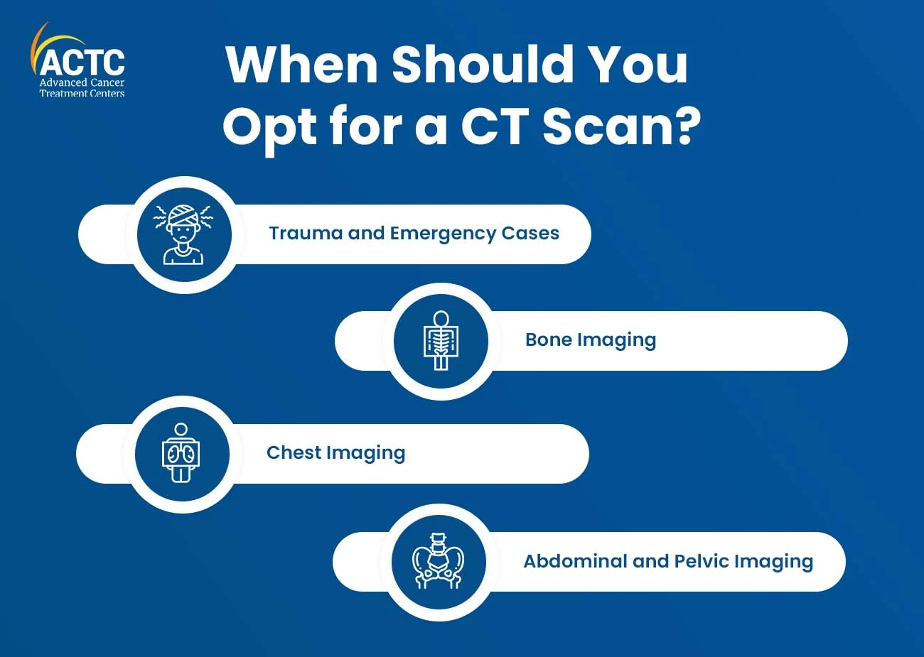
While they are effective for imaging many parts of the body, they are particularly useful for:
Trauma and Emergency Cases
Bone Imaging
Chest Imaging
Abdominal and Pelvic Imaging
One of the significant advantages of MRI is that it does not use ionizing radiation, making it a safer option for repeated imaging studies, particularly for vulnerable populations such as pregnant women and children.
MRI excels in providing detailed images of soft tissues, making it indispensable for the evaluation of the brain, spinal cord, and musculoskeletal system.
MRI allows imaging in multiple planes (axial, sagittal, and coronal), providing a comprehensive view of the anatomy from different perspectives.
Functional MRI (fMRI) is a specialized technique that measures and maps brain activity, offering insights into neurological functions.
Certain MRI exams require the use of contrast agents, which can cause allergic reactions in some individuals. Additionally, there are concerns about the safety of certain contrast agents, especially in patients with impaired kidney function.
The enclosed nature of traditional MRI machines can trigger claustrophobia in some patients, potentially affecting image quality if they struggle during the procedure.
MRI scans tend to take longer than CT scans, which may be challenging for patients who find it difficult to remain still for extended periods.
CT scans are known for their speed, making them ideal for emergencies where quick diagnosis is crucial.
CT scans provide exceptional images of bones and are particularly effective in identifying fractures and abnormalities in the skeletal system.
They can also be used to image a wide range of body parts, making CT scans a versatile tool in diagnostic imaging.
CT scans offer high spatial resolution, producing detailed images that are useful for assessing small structures and abnormalities.
The main drawback of CT scans is the use of ionizing radiation, which poses potential risks, especially with repeated or unnecessary scans.
While CT scans are excellent for imaging bones, they provide less detailed images of soft tissues compared to MRI.
Similar to MRI, some CT exams may require the use of contrast agents, which can lead to allergic reactions and carry risks for patients with impaired kidney function.
CT scans may not be the preferred imaging modality for certain conditions, such as evaluating joint and soft tissue injuries, where MRI offers superior contrast.
For pregnant individuals, the choice between MRI and CT scans depends on the medical necessity and the specific condition being evaluated. Generally, MRI is considered safer during pregnancy due to its lack of ionizing radiation
Pediatric imaging requires special consideration, and the choice between MRI and CT scan depends on different factors. MRI is often preferred for imaging the brain and musculoskeletal system in children due to its lack of ionizing radiation.
Patients with claustrophobia may find the enclosed space of a traditional MRI machine challenging. In such cases, CT scans, with their relatively quick imaging times, maybe more tolerable for claustrophobic individuals.
Both MRI and CT scans may involve the use of contrast agents, and patients with allergies or impaired kidney function need careful consideration. Medical professionals assess the individual's medical history and may opt for alternative imaging methods or adjust the contrast agent type and dosage accordingly.
MRI and CT scans are foundations in the constantly changing field of medical diagnostics, each having special advantages and uses. The medical condition being assessed, the required amount of detail, patient concerns, and the urgency of the diagnosis all play a role in which imaging modality is chosen.
The field of medical imaging has a bright future ahead of it as technology develops. From ultra-high field MRI to AI-driven image interpretation, these innovations aim to further improve diagnostic accuracy, patient outcomes, and overall healthcare delivery.
To learn more about the right fit for you, connect with the expert physicians at ACTC. Visit our website to book an appointment or call 352-345-4565
Since MRIs do not use ionization technology, they reduce the risk of developing cancer.
An MRI scan uses powerful magnets and radio waves to develop the images. CT scans on the other hand make use of ionization radiation.
In general, contrast resolution is better with MRIs, but spatial resolution is better with CT scans. This means that CT scans are useful for identifying boundaries, or the points at which one structure finishes and another begins.


January 07, 2026
A chemo port is a small device placed under your skin that makes recei...
KNOW MORE
December 24, 2025
It's natural to wonder if testosterone replacement therapy (TRT) is sa...
KNOW MORE

December 24, 2025
A rash that will not calm down is scary, especially when it changes or...
KNOW MORE
December 24, 2025
Florida’s lung cancer burden remains significant and affects many fa...
KNOW MORE
December 24, 2025
A partial hysterectomy, also called a supracervical hysterectomy, is s...
KNOW MORE

December 24, 2025
Finding a rash on your breast can be unsettling, but remember, many ra...
KNOW MORE
