
Book a Consultation
Thank you!
Your form has been sent successfully.

Meninges are protective tissue layers that surround the brain and spinal cord and help to circulate cerebrospinal fluid that cushions the brain from injury and provides nutrition to the brain and spinal cord. According to the American Cancer Society, meningioma is the most common primary brain tumor that develops in the meninges. Meningioma is a slow-growing tumor that may put pressure on the brain or spinal cord. Meningioma may be cancerous (malignant) or noncancerous (benign).
Based on the origin of the cancerous cells, meningioma types can be classified as
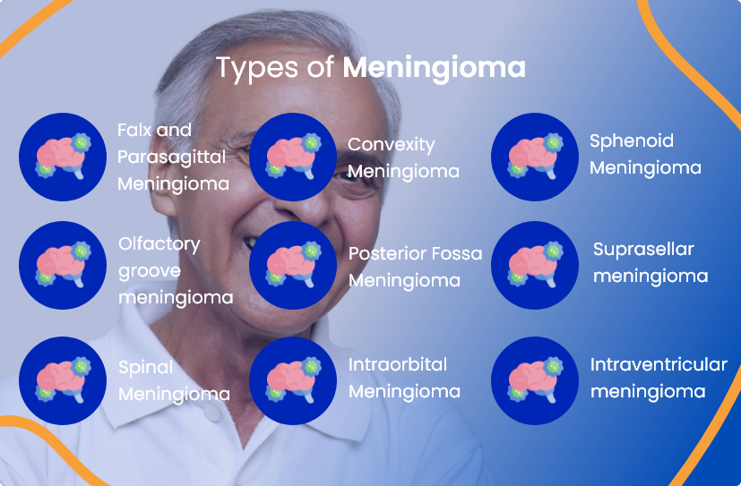
These are the most common meningiomas that account for approximately 25% of all cases. Falx meningioma occurs in the brain membrane present in the groove between the left and right sides of the brain (falx membrane). Parasagittal meningioma develops at the top of the falx membrane inside the skull.
Convexity meningioma accounts for about 20% of meningioma. It develops on the outer side of the brain.
Sphenoid meningioma occurs in the cells of the sphenoidal ridge that is located behind the eyes.
Olfactory groove meningioma occurs in the tissue layer that connects the brain to the nose.
Posterior fossa meningioma occurs in the cells at the back of the brain.
It develops in the cells located at the base of the skull, where the pituitary gland is located.
It manifests in the cells surrounding the spine at chest level and may press against the spinal cord.
It occurs in and around the cells of the eye sockets.
It develops in the chambers of the brain that carry cerebrospinal fluid throughout the brain.
Common signs and symptoms of meningioma may include:
 Seizures
 Headaches
 Blurred vision
 Personality or memory changes
 Unexplained nausea or vomiting
| Meningioma Type | Symptoms of Specific Meningioma |
|---|---|
| Falx and Parasagittal Meningioma | Leg weakness, headaches, seizures |
| Convexity Meningioma | Neurological complications like loss of coordination, muscle weakness, hearing loss, altered speech, personality changes, memory loss, and confusion |
| Sphenoid Meningioma | Numbness in the face, blurred vision, or loss of vision |
| Olfactory Groove Meningioma | Loss of smell, double/blurred vision, or loss of peripheral vision |
| Posterior fossa meningioma | Loss of hearing, facial numbness, involuntary spasm of face muscle, difficulty in swallowing |
| Supraseller Meningioma | Blurred or double vision, or total loss of vision |
| Spinal Meningioma | Pain in the back, limbs, or chest, numbness and weakness in the extremities (arms/legs), difficulty in bowel movement or bladder functions |
| Intraorbital Meningioma | Bulging of eyes, loss of vision |
| Intraventricular Meningioma | Personality changes, memory loss and confusion, headaches, or dizziness |
The treatment options depend on the type and stage of meningioma and the overall health of the patient.
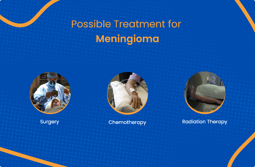
Surgery is considered as primary treatment for meningioma. A craniotomy is a surgical procedure where a skull piece is temporarily removed to allow meningioma doctors to access the brain and surgically remove the tumor and some surrounding healthy tissues. The extent of surgical removal depends on the size and spread of the cancer.
Radiation therapy uses high ionizing rays to destroy cancer cells that are otherwise inaccessible by surgery. Radiation can be delivered via an external machine outside of the body, called external beam radiotherapy. External beam radiation is the most commonly used radiotherapy to treat meningioma disease. Various types of external-beam radiotherapy used to treat meningioma are:
Chemotherapy uses anti-cancer drugs injected intravenously or orally, which enter the bloodstream and reach and destroy rapidly growing cancer cells.
The following tests and procedures may be recommended, depending on the symptoms of meningioma, age, and general health of the patient:

This exam includes checking various neuro functions like sight, hearing, reflexes, balance, and coordination. Abnormality in one or many functions may suggest which brain areas are affected by the tumor.
CT or MRI scans usually guide stereotactic needle biopsy to reach inaccessible brain areas. A meningioma doctor drills a small hole in the skull and inserts a needle to remove the tissue, which is later microscopically investigated for meningioma. The biopsy in meningioma may also be done postoperatively (after the complete removal of the tumor) to detect the presence of cancerous cells.
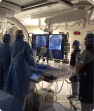
A cerebral angiogram is a diagnostic procedure where a catheter that is inserted into the arm/groin is guided to the brain area. A series of X-rays are taken after the contrast dye is injected into the catheter to examine the blood vessels of the brain. Contrast dye provides a detailed view of the blood vessels that circulate blood in the brain. A cerebral angiogram may detect a blocked vein or abnormal blood vessels that may be feeding the tumor.
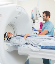
High-intensity radiation is used to produce detailed 3-dimensional computerized images. They are used to locate and measure the tumor's size. Various imaging tests used are:
The cancer specialists at ACTC in Florida offer outstanding patient care by prescribing personalized and evidence-based treatment plans tailored to individual patients' needs. We aim to foster a positive environment that focuses on physical and mental health throughout a cancer patient's journey.
The following are our providers who you can consult at ACTC:

MD, Hematology & Oncology

MD, Ph.D., Hematology/ Medical Oncology

MD, Radiation Oncologist

If you or a loved one has been diagnosed with meningioma, a detailed discussion with your primary physician will help you understand your condition better. Your primary care doctor can then refer you to an advanced specialty center, such as ACTC in Florida.
As one of Florida's best cancer centers, we understand how a cancer diagnosis and therapy impact a person's physical and emotional well-being. Therefore, we work hard to make patients and their families feel secure. We understand how imperative it is for you and your loved one to make informed choices and play an active part in your medical care. We at ACTC strive to support you at every step of diagnosis, staging, treatment, and long-term follow-up in one convenient location. Our cancer specialists are backed up by qualified clinical staff with over two decades of experience and a reputation for providing personalized meningioma treatment.
Schedule a consultation by calling
 352-345-4565
According to the American Cancer Society, various factors that increase the risk of meningioma include genetic factors, previous history of cancer, and a weakened immune system. The risk increases with age, and women are more commonly affected than men.
Meningioma is less likely to recur as compared to other brain tumors. Most meningiomas are benign and may be cured by complete surgical resection.
There are several factors that determine the prognosis (chances of recovery) of meningioma disease. They include the patient's general health and age, location of the tumor, metastasis of the tumor, type and grade of tumor, or if cancer has recurred.
Schedule a consultation by calling
 352-345-4565